
Organ Science
Key takeaways
- Ding et al. built aging models for more than 40 cell types from plasma proteins measured in 60,542 people across three cohorts, finding that 20–25% of individuals showed accelerated aging in a single cell type that an organ-level score would have averaged away.
- Individuals carrying two APOE4 alleles with extreme astrocyte aging signatures had triple the risk of incident Alzheimer's disease; those with youthful astrocyte signatures had reduced risk—associations that held after accounting for APOE genotype at the organ level.
- A comparative proteogenomic analysis found fewer than 30% of protein quantitative trait loci are shared between cerebrospinal fluid and plasma, meaning brain cell-type clock signals in blood are plasma proteomic signatures enriched for cell-type-associated proteins, not direct readouts of brain cell biology.
- No published intervention study has demonstrated that plasma-proteomic cell-type aging scores can be lowered; the measurement science is established at population scale, but the intervention literature does not yet exist.
For three years, the leading question in plasma proteomics and aging was which organs were aging faster than the rest. A study published in Nature Medicine in June 2026 answers a harder question: which cell types within those organs. Ding et al. analyzed more than 7,000 plasma proteins in 60,542 people across three cohorts and built machine-learning models to estimate the biological age of more than 40 cell types, including neurons, astrocytes, immune cells, and endocrine cells. Those cellular aging signatures predicted incident disease and mortality over 15 years of follow-up. The resolution is genuinely new. So are the interpretive limits.
What just changed
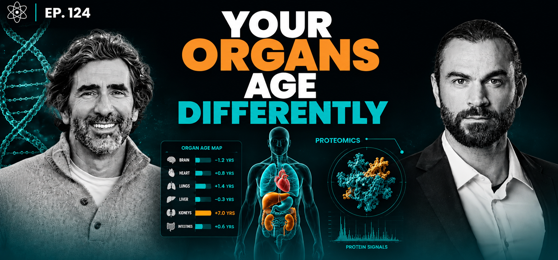
An organ clock asks: is your heart aging faster than the rest of you? A Cell Clock asks: which cells inside that heart, or that brain, are driving it?
The distinction matters because organs are not homogeneous. The brain contains neurons, astrocytes, oligodendrocyte precursor cells, microglia, and more, and they do not age in lockstep. A single organ-level score averages across all of them. If your astrocytes are aging rapidly while your neurons are not, the organ score absorbs that difference and returns something closer to the mean. The cellular signal disappears into the aggregate.
What Ding et al. demonstrated is that plasma proteomics, measuring proteins circulating in blood at sufficient scale, can resolve those within-organ differences. The models they built estimate biological age for more than 40 cell types from a blood draw. That is the resolution upgrade: not organ versus whole body, but cell type versus organ.
These are plasma proteomic signatures statistically linked to specific cell-type biology. The proteins were selected because they are enriched for expression in particular cell populations, based on reference data. What the models measure is pattern in blood; what they infer is something about cellular aging. The distinction between those two things becomes more important the further the interpretation travels from the blood draw toward the brain.
What the paper found
Ding et al. drew on three cohorts: the Global Neurodegeneration Proteomics Consortium (14,281 participants), the 1946 National Survey of Health and Development (1,803 participants), and the UK Biobank (44,458 participants), for a total of 60,542 individuals with up to 15 years of follow-up. More than 7,000 plasma proteins were analyzed using two separate measurement platforms: SomaScan (7,289 proteins) and Olink (2,923 proteins). Convergent results across both platforms strengthen confidence in the signals, though the platforms are not interchangeable and results should not be pooled without adjustment.
The headline population finding: 20 to 25 percent of individuals showed accelerated aging in a single cell type. Between 1 and 3 percent showed accelerated aging in ten or more cell types. A whole-organ score would have averaged the first group's signal away entirely.
The APOE4 and astrocyte finding is the sharpest clinical illustration. Individuals carrying two copies of the APOE4 allele who also showed extreme astrocyte aging had triple the risk of incident Alzheimer's disease. Those with two copies of APOE4 but youthful astrocyte aging signatures had substantially reduced risk. These associations held after accounting for APOE genotype at the organ level, meaning the cell-type signal carried information beyond what genetics alone explained.
Independent replication of the organ-level foundation is worth noting. Wang et al., in Nature Aging 2026 (n=43,616, UK Biobank, validated in China Kadoorie Biobank n=3,977 and Nurses' Health Study n=800), found cross-cohort correlations of r=0.93 to 0.98 for organ-level proteomic aging clocks. That figure applies to organ clocks. Cell Clock cross-method concordance at cellular resolution is not yet established in the published literature. The replication gives the platform credibility; it does not transfer automatically to the finer-grained models.
The interpretive limit
A high score on the astrocyte clock means the plasma proteomic pattern associated with older astrocyte biology is elevated in that individual's blood. It does not mean the researchers directly sampled astrocytes, or that the proteins measured came only from astrocytes, or that the signal is a transparent readout of what is happening inside brain cells.
This distinction becomes pointed when you consider the relationship between plasma and cerebrospinal fluid. A comparative proteogenomic analysis published around the same time as Ding et al. found that fewer than 30 percent of protein quantitative trait loci are shared between CSF and plasma (Western, Cruchaga et al., May 2026). The two compartments have substantially different regulatory environments, and brain-specific biology is not simply diffusing intact into peripheral blood.
Field commentary at Alzforum raised precisely this point: the "astrocyte aging," "OPC aging," and "inhibitory neuron aging" signals are more accurately described as plasma proteomic signatures enriched for proteins associated with those cell types, not direct measurements of the biological age of the corresponding brain cells. That is not a reason to dismiss the empirical associations. Ding et al.'s predictions hold in the data: the signals predicted disease and mortality over 15 years and showed a biologically coherent relationship with APOE4 status. Those are real findings. What remains open is the mechanism. Why does a plasma protein pattern linked to astrocyte biology predict Alzheimer's risk? Do astrocyte-derived proteins cross the blood-brain barrier in informative quantities? Does astrocyte dysfunction produce downstream systemic effects detectable in blood? The data support the association. They do not yet explain it.
The precision-medicine promise of Cell Clocks rests partly on the assumption that a plasma signal labeled "neuronal aging" reflects what is happening inside neurons. The CSF-plasma concordance literature is the honest counterweight to that assumption.
Where this leaves us
No published intervention study has demonstrated that plasma-proteomic cell-type aging can be lowered. One small exercise study involving 26 sedentary men over 12 weeks, with no described control arm, moved an organ-level clock by approximately 10 months. That is a signal worth investigating, not a finding worth concluding from. At the cell-type level, the intervention literature does not yet exist.
This is not an unusual position for a fast-moving field. The measurement science is ahead of the intervention science, which is exactly where it should be. You cannot design an intervention study around a biological endpoint until you have a reliable way to measure that endpoint. Ding et al. establishes that cell-type-specific aging can be estimated from plasma proteomics at population scale and that those estimates predict meaningful outcomes. That is the prerequisite for asking whether the estimates can be changed.
The questions that follow are the consequential ones. How consistently does an individual's cell-type aging score reproduce on retest? Which cell types show the most modification-responsive signals in observational data? Does lowering an astrocyte aging score in blood translate to reduced Alzheimer's incidence, or does the association only run in one direction? The answers require longitudinal studies with intervention arms and predefined endpoints. They are not yet in the literature.
What Ding et al. has done is sharpen the target. Twenty to twenty-five percent of people show accelerated aging in a single cell type that a whole-organ score would average away, and that cellular signal predicts disease and death over fifteen years, from a blood draw. The resolution is real. The work of establishing what to do with it has started.
Sources: Ding DY et al. Plasma proteomic signatures of cellular aging predict human disease. Nat Med 32:2060–2072 (2026). Wang et al. Nat Aging 6:162–180 (2026). Western, Cruchaga et al. proteogenomic CSF-plasma comparative analysis, May 2026 (cited in Alzforum commentary on Ding et al.). Alzforum expert commentary on Ding et al.: alzforum.org/papers/plasma-proteomic-signatures-cellular-aging-predict-human-disease. Oh et al. Nature Med 2025;31(8):2703–2711. Oh et al. Nature 2023;624:164–172.
Early access
Discover your OrganAge™
Join our waitlist to get priority access to OrganAge™ and Vero Compass™ as they become more widely available.
Get early accessEarly access
Discover your OrganAge™
Join our waitlist to get priority access to OrganAge™ and Vero Compass™ as they become more widely available.
Get early access